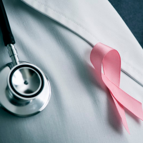
Mammograms save lives, and McLaren Greater Lansing's Breast Care Center is using the latest technology to detect breast cancer earlier, allowing for more effective treatment.
Tomosynthesis 3D imaging, also known as 3D mammography, is a screening and diagnostic tool producing images as small as one millimeter thick. The technology is able to create both 3D and standard 2D images that allow expert radiologists to look for breast cancer.
The only difference between a 3D and 2D mammogram is the technician will take images from a variety of angles to create a 3D image of the breast. In a standard mammogram, both dense breast tissue and cancer appear white, which makes breast cancer harder to detect. On average, the 3D mammogram finds 20 to 65 percent more invasive cancers than conventional mammography alone.
People with dense breast tissue, a personal history of cancer, and who are between the ages of 40 and 60 can benefit most from 3D mammography. The use of 3D imaging reduces unnecessary additional imaging from a screening exam by up to 40 percent, and the technology is also used in symptomatic patients.
"Something as simple as getting regular mammograms can save your life," said Jesse Amezaga, M.D., Radiologist with McLaren Greater Lansing's Breast Care Center. "With 3D mammography technology, our experts can more accurately detect and diagnose breast cancer."
The American College of Radiology has designated the McLaren Breast Care Center as a Breast Center of Excellence, and McLaren Greater Lansing is the first and longest-running mid-Michigan hospital to receive national accreditation for its breast care program by the National Accreditation Program for Breast Centers (NAPBC).